
Farm Tours
We offer farm tours for smaller groups and families to meet, feed, interact with and walk with our alpacas.
For full details and to book your experience of these wonderful animals, please go to our reservation form.
It is very important that alpaca owners know the normal behaviours of each of their animals. Alpacas are stoic, meaning that they will try to hide the symptoms of any injury or illness so knowing what is normal makes diagnosis of illness or injury far easier. Sudden and rapid weight loss is often indicative of health issues so condition scoring or weighing your alpacas on a regular basis is valuable. Visual clues such as lack of energy, spending more time recumbent and reluctance to stand can indicate illness.
Please note: this information is provided only as a guide. If an owner cannot quickly determine the cause of any abnormal symptoms or behaviour by an animal, veterinary assistance must be sought.
This page covers the following internal and external parasites. Click below to go to the section of interest.
The parasitic nematode worm (Haemonchus contortus) infects the C3/abomasum stomach compartment where it attaches and sucks blood from the lining. The female BPW produces thousands of eggs every day which are expelled in the alpaca’s faeces onto the paddock where they hatch, develop into larvae and are ingested during grazing. They then pass into the stomach of the new animal and attach to the stomach wall, thus the life cycle is complete. Haemonchus eggs can survive for long periods on the pasture during dry weather. If alpaca dung middens are not removed, large numbers of eggs can build up. Warm wet weather (especially in March and April) will cause the eggs to hatch and allow infection or reinfection of an alpaca.
Treatment involves immediate oral drenching of the animal or an injectable wormer. Although conventional treatment is by routine drenching, there is now evidence of resistance to the drugs commonly used [25], a problem being seen worldwide. One approach now used is to drench only when an alpaca is shown to have Haemonchus eggs (or a burden of other worm eggs) is its faeces. Dectomax (Doramectin) given subcutaneously (SQ) is the usual choice although there is some discussion over the appropriate dosage for alpacas. Oral drenches such as Matrix, a combination of abamectin, levamisole and oxfendazole can alternatively be used should resistance be encountered. Note that care must be taken with any drench containing levamisole as it has a low safety margin at normal dosages. Accurately weighing animals and calculating the correct dose is the best approach here.
Liver flukes are parasitic flatworms that infect the liver and bile ducts of alpacas, most commonly in animals grazing on wet or swampy ground where snail intermediate hosts are present.
Alpacas are most likely to be affected by liver flukes when they live on swampy ground which harbour snails. Fascioliasis is a condition that affects many animal species worldwide. The usual fluke associated with alpaca infections is Fasciola hepatica, a primitive animal with a complex life cycle.
Adult flukes live in the bile duct where they shed eggs into the intestine. These eggs fall onto the paddock along with the faeces. Simply, those falling into water will hatch into a miracidium which seeks out and bores into a snail where it develops into a cercarium. These leave the snail and attach themselves to plant leaves as a cyst which are ingested by grazing animals. Once in the gut, the immature flukes hatch from the cysts, burrow through the intestinal wall and migrate to the liver, thus the life cycle is complete. For those interested, there is an excellent YouTube video showing the entire life cycle using time-lapse photography.
Two forms of fascioliasis are recognised, acute and chronic. The acute form is the result of a massive intake of cysts and produces symptoms in alpacas of weight loss, depression and a refusal to stand - all a result of liver damage. The chronic condition is caused by progressive damage to the bile ducts leading to the alpaca losing appetite, anaemic and reluctant to move. Death may result in the absence of treatment. A further complication of the chronic form is that conditions at the liver become suitable for certain Clostridial bacteria to grow - a situation that can be prevented by an annual 5-in-1 clostridial vaccine.
Diagnosis must be made by a vet. An enzyme-linked immunosorbent assay (ELISA) test is the best diagnostic tool and can detect antihepatica antibodies in blood serum. The most effective drug of choice against liver fluke is triclabendazole (e.g. Fasinex) which is toxic to all life stages of Fasciola and has a wide safety margin in sheep and cattle. This flukicide is off-label for alpacas but known to be effective.
Coccidia are microscopic protozoan parasites that infect the intestinal tract of alpacas, most commonly caused by species of the genus Eimeria.
In New Zealand, four Eimeria species are known to infect llamas and alpacas. Of these, Eimeria macusaniensis is considered the most clinically significant gastrointestinal pathogen in alpacas of all ages and is the species discussed here. Infections occur worldwide and the parasite is likely ubiquitous in the environment, meaning most animals are exposed at some point in their lives.
Mammalian coccidia are highly host-specific; alpaca Eimeria species are not infective to other domestic or wild ruminants, and vice versa. Infection begins when transformed cysts (oocysts) are ingested from contaminated pasture or feed. These cysts hatch in the gut, penetrate the intestinal wall, and undergo development, damaging the gut lining in the process.
This damage may result in diarrhoea and the shedding of large numbers of cysts back onto the paddock. The cysts are extremely resistant to environmental degradation and can survive many chemical and physical treatments. Once E. macusaniensis is established on pasture, decontamination is very difficult. Preventive management, including regular midden removal and treatment of infected animals, is therefore essential.
Most infections are asymptomatic and self-limiting. However, when parasite numbers are high, clinical coccidiosis can occur.
Clinical signs are non-specific and may include anorexia, weakness, weight loss, colic, and diarrhoea. Of particular importance is the long prepatent period of E. macusaniensis, which ranges from 33 to 42 days. Alpacas may suffer severe intestinal damage or death before cysts are detectable in faeces.
Diagnosis and treatment must be undertaken by a veterinarian. The drug of choice is oral toltrazuril (Baycox). Although E. macusaniensis has the potential to cause serious disease in alpacas of all ages, studies suggest its frequency as a sole pathogen may be overstated and that co-infection with other pathogens may occur [69]. An excellent review of coccidiosis in South American camelids is available [68].
Fly strike, also known as myiasis, is a condition in which blowfly larvae (maggots) infest damaged skin, causing rapid tissue destruction and systemic illness.
Fly strike is well recognised as a serious condition in sheep and may occasionally affect alpacas, particularly during warm weather. Risk factors include short fleece, skin wounds, damp fleece, or contamination with faeces.
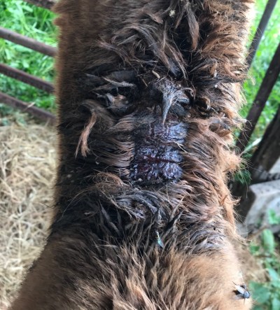
Blowflies lay eggs on damaged or soiled skin. These hatch within 12–24 hours, after which the maggots begin to degrade and liquefy underlying tissues. Toxins released from decomposing tissue and ammonia secreted by the larvae are absorbed into the bloodstream, resulting in systemic illness.
If untreated, fly strike can progress rapidly and may be fatal. Once established, the odour of tissue breakdown attracts additional flies, worsening the infestation. Early detection is therefore critical.
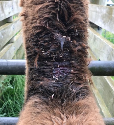
Regular inspection of the herd is essential, particularly after shearing. Increased numbers of flies around an animal should prompt immediate examination for wounds or skin abrasions.
Diagnosis is made by visual identification of maggots within affected areas. If immediate veterinary assistance is unavailable, the area may be lightly sprayed with a domestic fly spray to kill exposed larvae. Veterinary treatment remains essential and typically includes appropriate insecticides and antibiotics to control infection and prevent complications.
Mites and lice are external parasites that infest alpaca skin and fleece, potentially affecting animal welfare, fleece quality, and biosecurity.
Alpacas can be infected by mites of several genera causing specific skin conditions, Sarcoptes, Chorioptes, Bovicola and Psoroptes. The mange mite (Sarcoptes scabiei) is likely the most serious of these parasites and causes sarcoptic mange, otherwise known as scabies. Mite and lice infestations can impact alpaca welfare and fleece quality making early detection and treatment crucial.
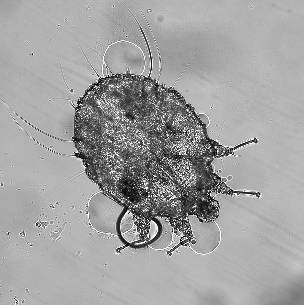
Photo by Arthur Goldstein - CC BY-SA 4.0
Sarcoptes. Sarcoptic mange occurs when the mite burrows into the skin of the less hair covered areas such as the legs, ears and belly and these develop bald spots, show flaking and crusting. The skin may become thickened as the disease develops and the alpaca shows intense itching (pruritus). Damage to the skin caused by scratching may become infected. Scabies is highly contagious and may be a significant zoonotic risk [20], i.e. it can spread to other animal species including humans.
Chorioptes mites are the commonest type of mite found on alpacas. They live the entire three weeks of their life cycle on the surface of the skin where they feed on dead cells. When caused by C. bovis, they are found particularly on the lower legs, feet and tail with symptoms of mild pruritus, alopecia and scaling of the feet and tail base resulting. Species identification is based on morphology (shape). Insecticidal sprays (eg. fipronil) are used in alpacas for eliminating Chorioptes mites as it kills them within two hours of contact and the treatment lasts up to one month although repeat treatments at three weeks are recommended [49].
Psoroptes mites can infect alpacas causing crusting weeping lesions which are more confined to the ears and as a result, the animal exhibits head shaking and scratching. The Psoroptes mite affecting alpacas may be the same species that causes sheep scab.
Bovicola (Lepikentron) breviceps lice are specific to alpacas and llamas and likely arrived in New Zealand around 2000. The louse feeds by chewing skin flakes, especially around the neck, shoulders and back though in low-grade infection, it does not appear to have any adverse effect on alpaca health or fleece quality [78]. If the animal is heavily infested, there can be mild to moderate itching, fleece damage and matting.
Prevention.
In all cases, skin scrapings can be taken from the affected areas and examined under the microscope. The morphology (shape) of any mites seen is used to diagnose the condition. Treatment of these mite conditions is usually by veterinary prescribed insecticides and it should be noted that all animals in the herd may need examination and treatment to prevent reinfection.
Ticks are blood-feeding external parasites belonging to the spider family that attach to the skin of alpacas and other livestock.
Ticks use their specialised mouth parts to penetrate the skin and suck blood which they use for their nutrition and reproduction. Although New Zealand has a number of native tick species that mainly infest birds, it is the imported Asian longhorned tick (Haemaphysalis longicornis) which infects livestock with cattle, sheep, deer, goats and alpacas all being susceptible. This species has been shown to carry and transmit pathogens (bacteria and viruses), one of which is associated with causing theileriosis in cattle. In other parts of the world, H. longicornis is known as a vector of animal and human diseases such as tick borne fever, Japanese (Oriental) spotted fever and Russian spring-summer encephalitis. Theileriosis (Theileria sp.), Lyme disease (Borrelia sp.) and Spotted fever (Ricketsia sp.) are not found as human diseases in New Zealand. A full profile description [81] of the Asian longhorned tick is available for New Zealand.
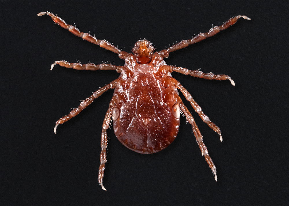
Before feeding, adult ticks are about 3 x 2mm wide and a red-brown colour (see picture). After feeding they will swell to almost 10mm long and 7mm wide and become blue-black. Although ticks are most visible attached to an animal, they actually spend under 20% of their lifespan on their host. After feeding, they will drop off and once in the grass will lay large numbers of eggs. These hatch, develop into larvae and then attach themselves onto a new animal; the life cycle is then complete. Given the time spent on the paddock, they are almost impossible to eradicate.
Ticks are most commonly found on the ears, legs and udder and frequently first seen when the restrained alpaca is being sheared. Any swollen ticks found should be carefully removed using a pair of fine tweezers - gripping the tick as close to the skin as possible and pulling the tick off. The tick should not be twisted or crushed when removing as this risks leaving the mouth parts still embedded and allowing an infection to start. The bite area should then be washed or sprayed with an antiseptic solution.
Affected animals may be treated using an avermectin group insecticide. Doramectin (Dectomax) has proven very effective [80] and is well tolerated in alpacas.
A new and highly effective treatment against ticks and mites has been recently reported [84]. Veterinarians used an oral preparation of fluralaner (sold as Flexolt in New Zealand) in an alpaca with sarcoptic mange and showed it to be a fast-acting treatment using a single dose of the drug.
Leptospirosis is a zoonotic bacterial disease caused by pathogenic species of Leptospira that can infect alpacas and many other mammals.
Leptospirosis is a
Leptospira bacteria are classified by their
Given that Leptospira requires water or high humidity to survive, they can be isolated from rivers, sewers, standing water sources, agricultural fields and moist soil.
Infection begins with Leptospires penetrating exposed mucous membranes or damaged skin. After a short incubation period, the bacteria invade multiple tissues including the liver and kidneys. Animals that are persistently infected will shed bacteria through the urine and this will be a source of infection. In alpacas, the disease is known for kidney and liver damage or failure. In pregnant females, abortion may occur or birth of a cria already infected.
Veterinary consultation is essentail as confirmation should be made from laboratory tests for serum antibodies levels in blood samples. Antimicrobial therapy will be needed and is usually effective. However, organ damage already caused by the infection can be permanent.
A vaccine is available in New Zealand (off-label for alpacas) which in addition to five clostridial species also covers Leptospira interrogans serovar Pomona, Leptospira borgpetersenii serovar Hardjo. This can be used in a similar way to 5-in-1 vaccines.
Most of the literature below can be accessed by clicking on the highlighted link. Some links will access the appropriate web page from which the article can be downloaded but others will immediately start downloading the full reference.
25. Waghorn, T.S., Leathwick, D.M., Rhodes, A.P., Lawrence, K.E., Jackson, R., Pomroy, W.E., West, D.M. and Moffat, J.R. (2006). Prevalence of anthelmintic resistance on sheep farms in New Zealand. N.Z. Vet. J. 54(6): 271-277.
26. Nisbet, A.J., Meeusen, E.N., González, J.F. and Piedrafita, D.M. (2016). Immunity to Haemonchus contortus and Vaccine Development. Adv. Parasitol. 93: 353-396.
47. Sanders, J., Xie, Y., Gazzola, D., Li, H., Abraham, A., Flanagan, K. Rus, F., Miller, M., Hu, Y., Guynn, S., Draper, A., Vakalapudi, S., Petersson, K.H., Zarlenga, D., Li, R.W., Urban, J.F., Ostroff, G.R., Zajac, A. and Aroian, R.V. (2020). A new paraprobiotic-based treatment for control of Haemonchus contortus in sheep. Int. J. Parasitol.: Drugs and Drug Resistance, 14: 230-236.
68. Dubey, J.P. (2018). A review of coccidiosis in South American camelids. Parasitol. Res., 117(7): 1999-2013.
69. McKenna, P.B. (2006). Eimeria macusaniensis in camelids - a brief review. Surveillance, 33(4): 8-10.
78. Palma, R.L., McKenna, P.B. and Aitken, P. (2006). Confirmation of the occurrence of the chewing louse Bovicola (Lepikentron) breviceps (Insecta: Phthiraptera: Trichodectidae) on alpacas (Lama pacos) in New Zealand. NZ Vet. J, 54(5): 253-254.
80. Doan, H.T., Noh, J.H., Kim, Y.H., Yoo, M.S., Reddy, K.E., Jung, S.C., Kang, S.W. (2013). The efficacy of avermectins (ivermectin, doramectin and abamectin) as treatments for infestation with the tick Haemaphysalis longicornis on rabbits in Korea. Vet. Parasitol., 198(3-4): 406-409.
81. Cane, R. (2010). Profile: Haemaphysalis longicornis Neumann, 1901. N.Z. J. Zool., 15: 585-586.
84. Sala, G., Gazzonis, A.L., Pravettoni, D., Cafiso, A., Grilli, G., Ferrulli, V., Boccardo, A., Di Cesare, F., Filippone Pavesi, L. and Zanzani, S. (2024). Effective treatment of sarcoptic mange in an alpaca (Vicugna pacos) using fluralaner: a case report. Vet. Res. Commun. 48: 1837-1843. https://doi.org/10.1007/s11259-024-10316-0.
We offer farm tours for smaller groups and families to meet, feed, interact with and walk with our alpacas.
For full details and to book your experience of these wonderful animals, please go to our reservation form.

Te Korito Alpacas now offer gift vouchers in a range of values — a perfect gift for animal lovers. Vouchers can be redeemed at our farm for a memorable alpaca experience or used toward unique goods in our farm shop.

During our farm tours, visitors ask many questions about the alpacas, from keeping them, on their health, to their breeding and much between. This page gives a selection of these along with concise answers and many pointers to more detailed information. This page is growing!
Directions - Once in Wanganui, drive up Somme Parade and when it becomes Papaiti Road (just past the cemetery), travel a further 7.4km alongside the river. We are the first house on the right with a large white letterbox and sign for the farm. On Google Maps we are shown as 'Te Korito Alpacas'.